
The question
Kidney abnormalities (cysts, stones, tumours) are diagnosed from CT volumes that are noisy, high-variance, and read under time pressure. Automated triage is attractive, but a false negative in this setting is not a metric; it is a missed tumour.
Two requirements therefore had to hold simultaneously: detection had to be reliable enough to be useful, and every prediction had to be auditable by a radiologist who was not involved in building the model.
Can a hybrid architecture capture both the local texture cues CNNs excel at and the global anatomical context radiologists use, while exposing the evidence behind each decision?
Why pure CNNs fall short here
Convolutions are local by construction. Stacking them enlarges the receptive field, but the inductive bias remains neighbourhood-centric: excellent for texture and edges, weaker for the long-range relationships that make a finding meaningful, where a lesion sits relative to the renal cortex, whether the contralateral kidney looks the same, how the structure relates to surrounding anatomy.
In noisy CT this shows up as false negatives: a lesion whose local texture is ambiguous but whose global context is clearly abnormal. Transformers have the opposite profile: global attention from the first layer, but data-hungry and weak on fine local detail when trained on modest medical datasets.
Architecture: TransConvNet
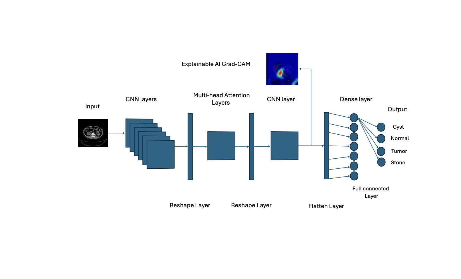
Rather than choose, TransConvNet composes them: convolutional stages extract local features with the sample efficiency CNNs bring, and transformer attention operates over those feature maps to model global dependencies.
Convolutional front end
Local feature extraction over CT slices (texture, edges, and lesion-scale structure), retaining the strong spatial prior that makes CNNs efficient on limited medical data.
Transformer attention stage
Self-attention across the extracted feature map, letting distant regions inform one another so anatomical context contributes to the decision.
Grad-CAM attribution head
Gradient-weighted class activation mapping produces a per-prediction heat map over the input, built into the delivered system rather than bolted on for the paper.
Explainability as a requirement
Grad-CAM computes the gradient of the predicted class score with respect to the final convolutional feature maps, weights each channel by its pooled gradient, and projects the result back to image space. The output is a heat map showing which regions moved the decision.
LcGrad-CAM = ReLU( Σk αck Ak ), αck = (1/Z) Σi Σj ∂yc / ∂Akij (1)
The practical value is not that it produces a pretty overlay. It is that a clinician can immediately spot the failure mode that metrics hide: a model that is right for the wrong reason: keying on a scanner artifact, a body-position cue, or a text marker burned into the image rather than the pathology itself.
Across representative slices the attribution concentrated on the renal region rather than on scanner borders, patient positioning, or annotation burn-in: the shortcut features that most often inflate medical benchmarks. That is the check worth running before trusting any accuracy figure on this kind of data.
Results
The hybrid outperformed the convolutional baselines it was compared against, with the gain concentrated, as hypothesised, in cases where local texture alone was ambiguous.
A near-perfect score on a curated benchmark says more about the dataset than the model. It indicates the task is well-posed and the classes separable under these conditions, it is not evidence of clinical readiness. The honest interpretation is that the architecture is sound enough to justify prospective evaluation, not that the problem is solved.
Limitations
- Single-source data. Trained and evaluated on one CT dataset. Scanner manufacturer, reconstruction kernel, and acquisition protocol all shift the input distribution: the same distribution-shift problem I pursue in the wireless work applies here and is untested.
- Slice-level, not patient-level. Clinical decisions are made over volumes and patient history, not isolated slices.
- Attribution is not explanation. Grad-CAM shows where the model looked, not why that region implies the class. It is a debugging instrument and a trust aid, not a causal account.
- No prospective validation. No reader study comparing model-assisted radiologists against unassisted ones: the evaluation that would actually establish clinical value.
What I'd do next
- Cross-scanner robustness. Apply the domain-adaptation machinery from my wireless work to multi-centre CT, and measure the degradation honestly.
- Volume-level modelling. Aggregate slice predictions with 3D context rather than treating slices independently.
- Calibration. Report confidence that means something: a well-calibrated model that abstains when uncertain is more useful clinically than a slightly more accurate one that never does.
- Reader study. Evaluate whether the attribution maps actually change clinician decisions, and in which direction.
Artifacts
BibTeX
@inproceedings{saeed2024transconvnet,
  author    = {Saeed, M. T. M. and Yousif, M. A. A. and Hakim, A. A. M.},
  title     = {TransConvNet: Enhancing Kidney Abnormality Detection in CT Imaging
               through Hybrid Transformer-CNN Model with Integrated Explainability},
  booktitle = {2024 12th International Scientific Conference on Computer Science (COMSCI)},
  address   = {Sozopol, Bulgaria},
  pages     = {1--5},
  year      = {2024},
  doi       = {10.1109/COMSCI63166.2024.10778503}
}